Advanced Labs Interpretation
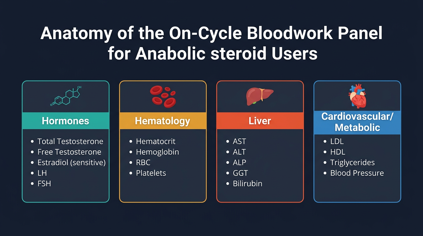
How to read a full blood panel in the context of active compound use, why reference ranges don’t apply, suppressed gonadotropins on cycle, hematocrit dynamics, advanced lipid markers, liver panel interpretation, IGF-1 confirmation for GH use, and thyroid monitoring.
- Interpret every major blood marker within its on-cycle context rather than population reference ranges
- Distinguish expected on-cycle findings from genuinely concerning results requiring action
- Analyse LH/FSH suppression in context and understand its implications for recovery
- Evaluate hematocrit and hemoglobin elevation and know when to intervene
- Interpret advanced lipid markers including ApoB in cardiovascular risk assessment
- Use IGF-1 testing to confirm GH axis response
- Assess thyroid markers in the context of GH and peptide use
Why Standard Reference Ranges Fail the On-Cycle User
Clinical laboratory reference ranges are derived from population studies of individuals who are, by definition, not using exogenous androgens. These ranges represent the 95th percentile interval of the reference population, they are descriptive statistics, not physiological targets. An on-cycle user operating with total testosterone at 3,000 ng/dL, hematocrit at 51%, and estradiol at 45 pg/mL will receive a lab report saturated with red flags on every reference range, while their actual clinical status may be entirely manageable.
The correct interpretive framework is not “am I within range” but rather “what does this value tell me about how this person’s physiology is currently functioning, and is there a concerning trajectory or threshold being approached?” This distinction requires knowing which values are expected consequences of deliberate compound use, which values represent dose-dependent effects that should be monitored rather than immediately treated, and which values represent genuinely concerning pathological signals regardless of context.
Advanced lab interpretation requires three things: a personal baseline (drawn pre-cycle), knowledge of what each compound class predictably does to each marker, and an understanding of the clinical thresholds at which intervention is indicated regardless of context. The sections below provide this framework marker by marker.
Testosterone and Androgen Markers On-Cycle
Total testosterone on an exogenous androgen cycle will be supraphysiological, this is expected and should not be acted upon in isolation. The relevant information is not the absolute value but whether it confirms the expected pharmacokinetic activity of the protocol. A total testosterone draw done at trough (just before the next injection) on 500 mg/week Testosterone Enanthate should produce a value of approximately 1,800–2,800 ng/dL depending on metabolic clearance rate, widely individual-variable. Values substantially below this might indicate underdosed product, unexpectedly rapid clearance, or a pre-trough draw timing error.
Free testosterone on cycle is elevated proportionally to total testosterone, modified by SHBG suppression from any co-administered compounds. If SHBG is markedly suppressed (by Winstrol, Anavar, or Mesterolone), free testosterone will be disproportionately elevated relative to total testosterone. This is not a problem per se, but high free testosterone means higher tissue androgen exposure per unit of total testosterone, which should inform expectations about androgenic sides.
SHBG may be dramatically suppressed on cycle (values below 10 nmol/L are common with oral compound use). Low SHBG per se does not require management, but it signals that the bioavailable hormone fraction is higher than total testosterone alone would suggest. The user whose SHBG is suppressed by Winstrol to 8 nmol/L is experiencing meaningfully higher androgenic tissue exposure than a user at the same total testosterone level with a normal SHBG of 30 nmol/L.
LH and FSH: Expected Suppression and Recovery Implications
On any meaningful exogenous androgen protocol, LH and FSH will be near-zero. This is physiologically expected, not pathological, the negative feedback loop that governs the HPTA is functioning exactly as designed in response to supraphysiological androgen levels. The pituitary’s response to high circulating androgens (and, via aromatization, high estrogens) is to reduce LH and FSH output, thereby reducing endogenous testicular testosterone and sperm production.
LH and FSH values near or at the laboratory detection limit during a cycle do indicate appropriate physiological response, not damage or pathology. The relevant question is not “why is LH suppressed” (the answer is always “because exogenous androgens are present”) but “will the HPTA recover after exogenous androgens are cleared?”
The markers that predict HPTA recovery quality are: the duration of the suppressive cycle (longer cycles produce deeper and more prolonged HPTA suppression), the compounds used (19-nor compounds, particularly at high doses and long durations, are associated with more prolonged suppression), the user’s age and pre-cycle baseline function, and genetic variation in hypothalamic-pituitary responsiveness. The clearest recovery indicator post-PCT is rising LH and FSH in the presence of rising total testosterone, this confirms hypothalamic and pituitary function is resuming. Persistent low LH/FSH at 8–12 weeks post-PCT with low total testosterone warrants endocrinological evaluation.
Hematocrit and Hemoglobin: The Erythropoietic Dose-Response
All androgens stimulate erythropoiesis, they increase red blood cell production by upregulating EPO secretion from the kidneys, directly stimulating bone marrow erythroid precursors, and suppressing hepcidin (a negative regulator of iron utilisation in red blood cell synthesis). Hematocrit elevation is therefore an expected finding on any androgen cycle, and its magnitude is broadly proportional to androgenic dose and duration.
The compounds with the greatest erythropoietic effect are, in approximate order: Testosterone (particularly at doses above 300 mg/week), Equipoise (disproportionately erythropoietic relative to its androgenic potency, one of its most notable properties at performance doses), Nandrolone (significant erythropoiesis at performance doses), and orally bioavailable 17-aa androgens to a lesser extent. Trenbolone also elevates hematocrit substantially despite its atypical receptor profile.
The clinical concern begins at hematocrit above 52%, blood viscosity at this level meaningfully increases thrombotic risk. Above 54%, the risk is clinically significant; above 55–56%, therapeutic phlebotomy is urgently indicated. Hematocrit should be monitored at minimum mid-cycle and the intervention threshold should be treated as non-negotiable. Therapeutic phlebotomy (removing 450–500 mL of whole blood, equivalent to a standard donation) acutely reduces hematocrit by 2–4 percentage points and is the only reliable acute intervention.
An important nuance: dehydration artificially elevates measured hematocrit. Draw conditions should be standardised, normal hydration status, no intense exercise within 24 hours. A hematocrit of 52% in a well-hydrated, rested state is more concerning than a 52% drawn after a hard training session in warm weather.
Advanced Lipid Analysis: Beyond the Basic Panel
The standard lipid panel (total cholesterol, HDL, LDL, triglycerides) is a starting point but does not capture the full cardiovascular risk picture in AAS users. Several advanced markers add meaningful resolution.
Apolipoprotein B (ApoB): Each LDL particle contains exactly one ApoB-100 molecule. ApoB therefore directly counts the number of LDL particles rather than measuring their total cholesterol content. A user with a standard LDL-C of 130 mg/dL could have this achieved by many small, dense LDL particles (each carrying less cholesterol, high particle count, high atherogenic risk) or fewer larger LDL particles (lower particle count, lower atherogenic risk). ApoB distinguishes these scenarios. In the context of androgen use, which shifts the LDL profile toward smaller, denser particles, ApoB is a more accurate cardiovascular risk marker than LDL-C alone. Target for cardiovascular risk reduction in AAS users: ApoB below 90 mg/dL is considered low risk; above 110 mg/dL with other risk factors warrants active intervention.
HDL functionality vs. HDL-C: Measured HDL cholesterol (HDL-C) reflects the total cholesterol transported within HDL particles but not the reverse cholesterol transport efficiency or HDL functionality. In androgen users, particularly those on 17-aa oral compounds, HDL can be structurally abnormal even when the total HDL-C is not catastrophically suppressed. This means HDL-C understates cardiovascular risk in oral androgen users, a limitation worth understanding when interpreting results.
Lipoprotein(a), Lp(a): An independent cardiovascular risk factor determined largely by genetics and elevated by androgens in genetically susceptible individuals. Worth including in a comprehensive baseline panel, particularly for users with family history of early cardiovascular disease. Elevated Lp(a) in the context of androgen-induced HDL suppression represents a compounding risk that should inform compound selection and cycle duration.
Fasting insulin and HOMA-IR: Insulin sensitivity is a key component of metabolic cardiovascular risk and is affected by androgens (variable effects at supraphysiological doses) and dramatically affected by MK-677 and exogenous hGH. Fasting insulin elevation above 10 µIU/mL in the context of normal fasting glucose warrants attention.
Liver Panel: ALT, AST, and Interpretation in Context
Liver enzymes elevation during oral compound use is expected, predictable, and ranges from clinically insignificant to immediately dangerous depending on magnitude and context.
ALT (alanine aminotransferase) is the most liver-specific of the standard hepatic markers, it is found predominantly in hepatocytes and its elevation reliably indicates hepatocellular stress or damage. AST (aspartate aminotransferase) is present in liver but also substantially in skeletal muscle, cardiac muscle, and red blood cells. In athletes performing intense resistance training, AST elevation of 2–3 times the upper limit of normal may reflect muscle damage rather than hepatic pathology, this is why ALT is the more reliable hepatic marker in trained individuals.
The interpretation framework: ALT 1–2 times ULN during an oral cycle is expected and manageable. ALT 2–3 times ULN warrants dose reduction and follow-up testing in 2–4 weeks. ALT above 3 times ULN requires cessation of the causative oral compound and reassessment at 4 weeks. ALT above 5 times ULN or any combination of ALT elevation with elevated bilirubin, jaundice, right upper quadrant pain, or dark urine constitutes a hepatic emergency requiring same-day medical evaluation.
TUDCA 500 mg/day during oral compound cycles provides genuine documented hepatoprotection. Its mechanism, mitochondrial stabilisation and anti-apoptotic hepatocyte protection, directly addresses the primary hepatotoxic mechanism of 17-aa compounds. Skipping this in a well-managed protocol is not an option.
GGT (gamma-glutamyl transferase) is a useful sensitivity marker, it tends to rise before ALT with hepatic stress and remains elevated after recovery. A trending rise in GGT over several weeks can serve as an early warning to reduce oral compound exposure before ALT becomes significantly elevated.
IGF-1 Testing for GH Users
IGF-1 is produced primarily in the liver in response to GH signaling and is the primary downstream mediator of GH’s anabolic effects. It is also the practical confirmation marker for GH axis activity, a GH dose that is not raising IGF-1 is not producing meaningful anabolic or body composition benefit, regardless of GH blood level.
For users of exogenous hGH, IGF-1 should be tested after 4–6 weeks of consistent dosing to confirm response. The target range in performance use is typically the upper quarter of the age-adjusted normal range, approximately 200–350 ng/mL for adults under 40 (ranges vary by laboratory and age). IGF-1 above 400–450 ng/mL at sustained doses raises concerns about long-term adverse effects on cell proliferation and should prompt dose reduction. IGF-1 that has not risen appreciably despite several weeks of supposedly adequate hGH dosing should prompt product quality investigation, underdosed or counterfeit GH is extremely common in the research chemical and grey-market pharmaceutical space.
For peptide secretagogue users (CJC-1295/Ipamorelin, MK-677), IGF-1 testing at 6–8 weeks of consistent use confirms that the protocol is producing the intended axis stimulation. Typical IGF-1 responses: MK-677 at 25 mg/day produces reliable 30–50% IGF-1 elevation from baseline; twice-daily CJC-1295 non-DAC + Ipamorelin protocols produce more variable responses (15–40% elevation) depending on compliance, timing relative to meals and sleep, and individual somatotroph responsiveness.
Thyroid Panel for GH and Peptide Users
The thyroid gland is connected to the GH axis through multiple feedback mechanisms. GH and IGF-1 stimulate peripheral T4 to T3 conversion, and thyroid hormones in turn influence GH secretion patterns. Subclinical hypothyroidism can attenuate GH response to secretagogues and reduce the efficacy of GH-axis manipulation strategies.
A baseline thyroid panel (TSH, free T3, free T4) is therefore particularly important for users of hGH, MK-677, and peptide secretagogues. TSH above 3.0 mIU/L combined with low-normal free T3 and T4 should be recognised as potentially impeding GH-axis protocols even if the clinical threshold for hypothyroidism treatment has not been formally met.
Long-term MK-677 use has been associated with modest TSH suppression in some users, possibly via elevated IGF-1's inhibitory effect on TSH secretion, a loop similar to the one seen with exogenous hGH at high doses. Persistent TSH suppression below 0.5 mIU/L with normal T3/T4 in an MK-677 user is worth monitoring but is typically not clinically actionable unless symptomatic. Any significant alteration in thyroid axis markers in the context of peptide or GH use should be discussed with an endocrinologist rather than self-managed.
Selected references for major clinical, mechanistic, or protocol claims. Community-practice points may not be cited individually.